Thursday, April 28, 2011

Vote RuBisCO: Practically Perfect!

Life requires carbon.
Every day, you need to eat about 250g of carbon to live.[1]

That may not sound like much, but to feed every human on the planet, plants have to provide 600 million metric tons of carbon each year! (And that’s if we were all vegetarians)

But don’t worry! The atmosphere contains over 2 billion metric tons of carbon right now, and we’re increasing that number every day![2]

There’s just one more catch: atmospheric carbon is CO2 – the fully oxidized form of carbon, which is totally useless to us.

Fortunately, there is a protein that can help! 

RuBisCO!
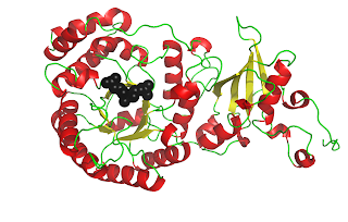
RuBisCO[3] catalyzes the carbon-fixing reaction in which a molecule of CO2 is added to the enediol form of ribulose-1,5-bisphosphate (RuBP), forming two molecules of 3-phospoglyceraldehyde (3-PGA). In these figures, you can see a crystal structure of the unactivated form of RuBisCO bound to its substrate RuBP[4] (above) and the activated form of RuBisCO bound to its two 3-PGA products[5] (below). Notice how the protein chain has undergone a conformational change in the activated complex, bringing the beta sheet behind the beta barrel. Also, activated RuBisCO contains a magnesium ion, shown in blue. These images show just one of the sixteen subunits of RuBisCO. For a better understanding of RuBisCO’s structure, see the “RuBisCO Images” post[6]

Let me give you four words to help you remember RuBisCO’s characteristics:
Pervasive: RuBisCO is the most abundant protein on the planet.
Perseverant: Day in and day out it, it works diligently to provide you with the carbon you need to live. Although RuBisCO can perform only three reactions a second, it keeps right on working, fixing carbon to give you delicious sweet corn, crisp cucumbers and juicy watermelon all summer long. 
Practically Perfect: Some have accused RuBisCO of substrate promiscuity, arguing that a more ideal protein would be able to better discriminate between CO2 and O2, but all efforts to improve this protein have failed! In fact, RuBisCO accomplishes a reaction that no chemist has ever accomplished: efficiently fixing CO2 straight from the atmosphere! This means it has to discriminate between CO2 and O2, two nearly featureless gases. Additionally, the transition state for fixing CO2 is higher energy than the transition state for fixing O2, which means that increasing the enzyme’s specificity for CO2 means decreasing its rate. Each RuBisCO is highly tuned to the CO2:O2 ratios and temperatures in its plant’s environment so that it balances specificity and speed. RuBisCO has not been improved because it is practically perfect!
Pervasive. Perseverant. Practically Perfect.
Show your appreciation to the diligent protein that fixes life. 
Vote RuBisCO

[1] My calculations based on recommended daily values for an average college student from http://www.healthcalculators.org/index.html  (313g carbohydrates, 85g protein, 69g fat) and percent carbon composition of carbohydrates (40%), proteins (~50%) and fats (75%).
[3] Information about RuBisCO was attained from the papers sited at http://rubiscofixeslife.blogspot.com/2011/03/all-about-rubisco.html.
[4] PDB file 1RCX
[5] PDB file 1AA1
Saturday, March 26, 2011

All About RuBisCo

“Distinct form I, II, III, and IV Rubisco proteins from the three kingdoms of life provide clues about Rubisco evollution and structure/function relationships”
Tabita et. al. in Journal of Experimental Botany, Vol. 59, No. 7, pp. 1515-1524, 2008

Rubisco is the most abundant protein in nature. It catalyzes the carbon-fixing reaction in which a molecule of CO2 is added to the enediol form of ribulose-1,5-bisphosphate (RuBP). The active site is located between two large subunits, and it involves residues from the amino terminus of one subunit and the carboxy terminus of the other. This site accepts both CO2 and O2, and its ability to distinguish between the two is limited. Tabita et. al. provide this figure to illustrate the competing reactions. Rubisco is found in four forms, and form I (consisting of eight large catalytic subunits and eight smaller subunits, annotated L8S8) is the most common, being found in eukaryotes and bacteria. However, the three other forms are not insignificant, and Rubisco-like protein shows a great deal of homology to the Rubisco proteins, and yet is not able to catalyze either carboxylation or oxygenation. The paper goes on to explore the evolutionary relationships between these protein forms and concludes that they are related by divergent evolution. Each form of Rubisco is optimized to suit the specific temperature and CO2/O2 ratio of the organism. 

“Despite slow catalysis and confused substrate specificity, all ribulose bisphosphate carboxylases may be nearly perfectly optimized”
Tcherkez et. al. in PNAS, Vol. 103, No. 19, pp. 7246-7251, May 9, 2006

Each cell invests large amounts of nitrogen into producing many Rubisco complexes; however, these complexes are both slow and nonspecific (unable to discriminate between CO2 and O2). Although some research groups have tried to improve upon Rubisco, they have only met with modest success. This paper shows that in fact, the slow catalysis is the enzyme’s way of discriminating between the two featureless gases. By making the transition state very similar to the products rather than the reactants, the enzyme can tell the difference between CO2 and O2; however, a product-like transition state is more ordered and makes the product bind tightly such that it decreases the catalytic rate. Thus, the enzyme must strike a balance between specificity and speed, and each individual Rubisco is optimized for the CO2/O2 ratio in which it is found: enzymes present in environments with lower CO2/O2 ratios have higher specificity and slower rates, while enzymes in environments with higher ratios may be more promiscuous and have faster rates. Thus, Rubisco cannot be dramatically improved because it is already nearly perfect.

“Highly Conserved Small Subunit Residues Influence Rubisco Large Subunit Catalysis”
Genkov and Spreitzer in the Journal of Biological Chemistry, Vol. 284, No. 44, pp. 30105-30112, October 30, 2009

Although the active site of Rubisco is found between the carboxyl-terminal α/β-barrel of one large subunit and the amino terminus of its neighboring large subunit, extensive studies of the large subunit have not yielded any significant increases in the protein’s catalytic rate or its specificity for CO2 over O2. Although the CO2/O2 specificity (termed Ω) differs between prokaryotes and eukaryotes, the large subunit is highly conserved; instead, the small subunit displays the most variation. These authors hypothesize that the small subunit may be more responsible for modulating Ω and set out to study the regulatory role of the small subunit. They replaced each of the ten residues which are highly conserved among all small subunits with alanine and monitored the effect on catalysis and specificity. None of the tested residues are essential for the Rubisco structural integrity or function. Three of the mutations showed no changes at all; one decreased the stability of the enzyme complex; two decreased the specificity of the enzyme; and four affected catalysis with differing changes in enzyme stability. Because the residues tested were conserved across all L8S8 Rubisco enzymes, these differences can only be used to explain the differences between L8S8 and L2 enzymes, but not the differences within the L8S8 subset.

Wednesday, March 2, 2011

Rubisco Images


RuBisCo, or ribulose-1,5-bisphosphate carboxylase oxygenase has 16 chains - 8 large chains and 8 small chains. Each large chain is paired with a small chain, forming a heterodimer. The heterodimers further dimerize, forming four-chain complexes which are axial symmetrical. Four identical tetramers circle up to form the overall protein. Above, the protein is color coded by chain. The long chains are green or blue and the short chains are yellow or red. Each quadrant (blue or green) has two large chains. The small chains fit above (toward the viewer) or below (away from the viewer, on the back side) the large chains, and they are slightly offset, sitting in between the large chain subunits.
To further explore the structure, the image above shows just one of the tetrameric subunits: two large chains and two small chains. In this image, the large chains are the darker shade and the small chains are the lighter shade. One large-small chain dimer is red, while the other is blue. This view is from the side of the protein, and the viewer is on the outside of the protein looking in. The spheres are ribulose-1,5-bisphosphate, the substrate of the protein.
Breaking down the structure further, a large-small chain dimer is shown above. This view is from the side of the large chain which normally faces the other long chain in the tetrameric subunit. The beta barrel with corresponding alpha helices surrounding it is visible under the substrate. The right-most portion of the structure contains a beta sheet. These structural features are portrayed more clearly in the image of the long chain below.
Because this protein is so large and complex, it does not lend itself well to more complicated images, for example, stick or line images displaying every amino acid, or mesh images displaying the contours of the surface, both of which are pictured below. These views mimic the first image in this post. The viewer is above the protein, looking down through the center of it.

Tuesday, March 1, 2011